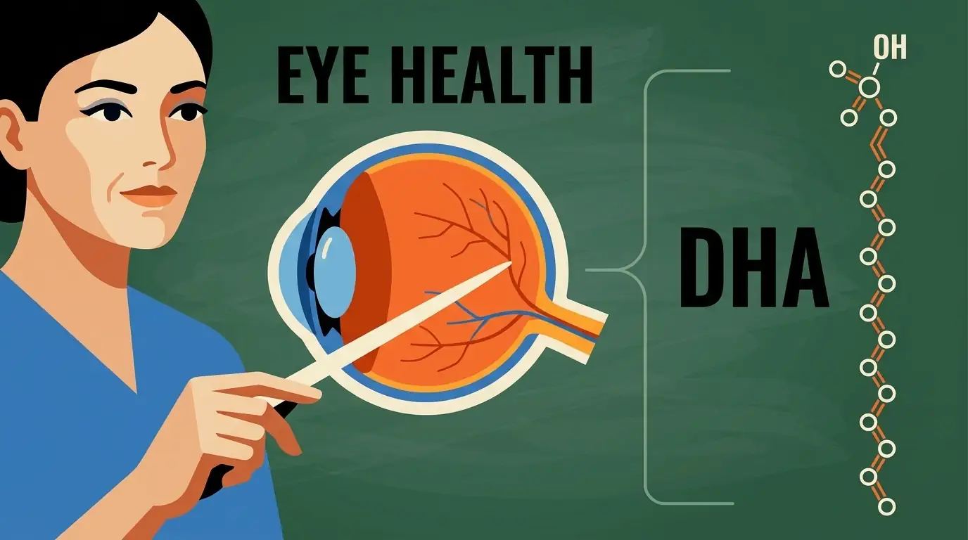
Title: The critical importance of DHA in eye health
Published: - Updated:
Author: Redacción Brudylab
Reviewer: Dra. Leia Garrote - Medical Director
Docosahexaenoic acid (DHA) is the main structural fatty acid of the retina, where it represents more than 50% of the membrane lipids in photoreceptors. Its role extends beyond nutrition: it is a precursor of neuroprotective molecules such as NPD1 and a key factor in the prevention of age-related macular degeneration. This article reviews its biology, dietary sources, and synergistic action with carotenoids and antioxidants.
The critical importance of DHA in eye health
The Critical Importance of Docosahexaenoic Acid (DHA) in Eye Health and Retinal Integrity
Omega-3 and eye health: why DHA is key to vision
Visual health results from a complex interaction between genetic factors, aging, environmental exposure, and nutritional status. Among the nutrients that support biological function, omega-3 fatty acids, and in particular docosahexaenoic acid (DHA), occupy a central and irreplaceable place. DHA is not an optional dietary component: it is a primary structural constituent of ocular tissues, especially the retina, where its presence is necessary for the correct transduction of light into nerve impulses.
The relevance of DHA in modern ophthalmology has grown as clinical and epidemiological research has linked its intake to the prevention and management of degenerative diseases. It is estimated that by 2040 approximately 288 million people will suffer from some degree of Age-Related Macular Degeneration (AMD) [1], underscoring the need for evidence-based nutritional interventions.
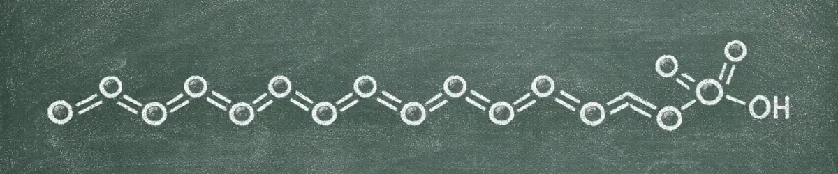
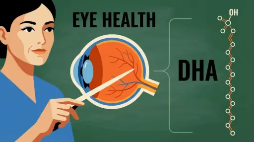
DHA is a long-chain polyunsaturated fatty acid with 22 carbon atoms and 6 double bonds (C22:6n-3). It is selectively concentrated in the central nervous system and the retina, a distribution that reflects the need for cell membranes to maintain extreme fluidity to facilitate the molecular processes of vision. As a tissue with exceptionally high oxygen consumption and constant exposure to light radiation, the retina continuously generates free radicals, requiring a specific lipid supply to support its function.
DHA: a structural component of the retina
The retina is a thin layer of nerve tissue that lines the back of the eyeball. Within this structure are the photoreceptors, specialized cells divided into rods (for vision in low light conditions) and cones (for visual acuity and color perception). DHA is the most abundant fatty acid in the membranes of these cells, making up between 50% and 60% of the total fatty acids in the outer segments of the rods [2].
The physics of fluidity and phototransduction
The high concentration of DHA in photoreceptors is not a static characteristic: it is an operational requirement for phototransduction, the biochemical process by which a photon of light is absorbed by rhodopsin, triggering a cascade of electrical signals. Thanks to its flexible molecular structure, DHA reduces the rigidity of the cell membrane and allows rhodopsin to change conformation at the speed required for vision.
La deficiencia de DHA altera las propiedades físicas de estas membranas, ralentizando la activación de la rodopsina y reduciendo la eficiencia del procesamiento visual. Esta alteración funcional se puede detectar clínicamente mediante electrorretinograma (ERG) como pérdida de agudeza visual y menor sensibilidad al contraste [3].
The recycling cycle and the Retinal Pigment Epithelium (RPE)
Photoreceptors are subject to continuous wear and tear: their outer segments shed and are phagocytosed by the Retinal Pigment Epithelium (RPE). The RPE does not destroy the DHA present in these cellular remnants, but actively recycles it, returning it to the photoreceptors for the synthesis of new discs. Even with this closed-loop system, a constant external supply is required to compensate for metabolic losses and accumulated oxidative stress [4].
| Retinal Layer / Cell | Role of DHA | Clinical Relevance |
|---|---|---|
| Photoreceptors (Rods) | Membrane fluidity for rhodopsin | Night vision and light sensitivity |
| Photoreceptors (Cones) | High-resolution vision support | Visual acuity and color perception |
| RPE (Reactive Pigment Epithelium) | DHA recycling and synthesis of protectants | Prevention of retinal atrophy |
| Synaptic Membranes | It facilitates neurotransmission | Integrity of the eye-brain nerve signal |
DHA and development of the visual system
The importance of DHA begins before birth. During the third trimester of pregnancy, there is a massive transfer of this fatty acid from the mother to the fetus through the placenta, coinciding with the accelerated formation of neuronal and retinal structures. DHA levels in early childhood are a direct predictor of future visual ability, including the maturation of the parvocellular pathway, responsible for processing fine details and colors [5].
DHA and maintenance of retinal integrity
As an individual ages, DHA shifts from being a driver of development to acting as a protective factor against two of the main enemies of vision: oxidative stress and chronic inflammation.
The discovery of Neuroprotectin D1 (NPD1)
One of the most relevant findings in ocular biochemistry was the identification of DHA as a precursor of Neuroprotectin D1 (NPD1). Under stress conditions, the RPE activates an enzyme that releases DHA from the membranes and transforms it into NPD1. This molecule acts on three fronts [6] [7]:
- It blocks cell death (apoptosis): it inhibits the activation of caspases and upregulates Bcl-2 family proteins that protect cell integrity.
- Controls inflammation: reduces the expression of pro-inflammatory genes that could damage the surrounding retinal tissue.
- It protects central vision: its synthesis is especially relevant in the macular area, where the density of photoreceptors is highest.
DHA and Age-Related Macular Degeneration (AMD)
Age-related macular degeneration (AMD) is the leading cause of irreversible vision loss in people over 60 in developed countries. It is characterized by the accumulation of deposits called drusen, degeneration of the retinal pigment epithelium (RPE), and progressive loss of photoreceptors. Epidemiological studies show that a higher dietary intake of omega-3 fatty acids is associated with a significantly lower risk of developing advanced forms of the disease [1]. DHA helps stabilize RPE membranes, improving their ability to clear metabolic waste and reducing the risk of choroidal neovascularization (the wet form of AMD).
| Protective Mechanism | Function of DHA / NPD1 | Outcome on Eye Health |
|---|---|---|
| Anti-apoptotic | Regulation of Bcl-2 and Bcl-xl proteins | It prevents the premature death of photoreceptors. |
| Anti-inflammatory | Inhibition of COX-2 and cytokines | It reduces collateral damage from chronic inflammation |
| Mitochondrial Stability | Preserves cellular energy | It maintains visual function under light stress. |
| Phagocytosis | Support the cleanup of waste in the EPR | It prevents the formation of druses and deposits. |
Why does the body need to obtain DHA through diet?
Although humans possess the enzymatic machinery to transform alpha-linolenic acid (ALA) into DHA, this process is a narrow and inefficient metabolic pathway. ALA is present in plant-based foods such as walnuts, flaxseeds, and chia seeds, but the conversion rate to DHA in healthy adults is generally less than 1% [8][9], and in many cases virtually undetectable. Since internal conversion cannot meet the demands of the retina and brain, DHA must be obtained directly from the diet or through supplementation.
| Nutrient | Typical Origin | Effectiveness for Eye Health |
|---|---|---|
| ALA | Flax seeds, chia seeds, walnuts | Low; minimal conversion to DHA (<1%) |
| DHA (Marine) | Oily fish, shellfish, seaweed | Maximum; it is incorporated directly into the retina |
| DHA (Supplement) | Fish oil or microalgae | High; allows controlled and purified doses |
Nutrition and eye health: the role of key molecules
Visual health relies on a group of molecules that work together. Research has identified that the combination of omega-3 fatty acids, carotenoids, and antioxidant vitamins offers the strongest protection against age-related vision decline.
Omega-3 fatty acids (DHA)
DHA is the only one of these nutrients with a structural function: it forms part of the physical components of photoreceptor membranes and is a precursor to survival signals such as NPD1. Its role is to ensure that the lipid substrate of the eye is flexible and resistant to chronic metabolic stress.
Macular carotenoids: Lutein and Zeaxanthin
Lutein and zeaxanthin are pigments that accumulate specifically in the macula, acting as a filter against high-energy blue light before it reaches the photoreceptors. They are estimated to be able to filter between 40% and 90% of this radiation [10]. Lutein is predominant in green leafy vegetables (spinach, kale), while zeaxanthin is more abundant in orange and yellow foods (corn, bell peppers, squash).
La distinción entre ambos carotenoides no es solo botánica: en la retina presentan distribuciones complementarias. La zeaxantina domina en el centro de la fóvea, donde la agudeza visual es máxima, mientras que la luteína predomina en la periferia macular. Esta distribución diferencial sugiere roles específicos que la suplementación combinada busca replicar.
Antioxidant vitamins (C and E) and Zinc
These molecules act on oxidative damage from different angles:
- Vitamin C: maintains the health of intraocular blood vessels and is associated with a lower risk of lens opacity.
- Vitamina E: Fat-soluble antioxidant that protects DHA fatty acid chains against lipid peroxidation, preserving the integrity of photoreceptor membranes.
- Zinc: an essential mineral for the metabolism of vitamin A in the retina and a cofactor of antioxidant enzymes.
The AREDS2 study established that the combination of lutein, zeaxanthin, vitamins C and E and zinc significantly reduces the risk of progression to advanced forms of AMD, and that replacing beta-carotene with lutein/zeaxanthin improves the safety profile, especially in smokers [1].
Frequently Asked Questions
What is the difference between DHA and other omega-3?
The omega-3 family includes three main members: ALA, EPA, and DHA. ALA is short-chain and is primarily used as an energy source. EPA has 20 carbons and plays a role in systemic inflammation and cardiovascular health. DHA has 22 carbons and is the only one that is massively concentrated in the brain and retina, forming part of their physical structure and supporting visual function.
Does the retina contain a lot of DHA?
Yes. The retina is one of the tissues with the highest concentration of DHA in the body. In the membranes of the photoreceptors where light is captured, DHA represents more than 50% of the fatty acids, a density that is not found in almost any other tissue except in specific areas of the brain [4].
Can DHA be produced from a plant-based diet?
The body can attempt to synthesize it from the ALA present in flax seeds or walnuts, but the conversion is extremely limited, generally less than 1% in healthy adults [9]. To maintain adequate levels in the retina, it is necessary to ingest preformed DHA directly from marine sources or supplements.
In which foods is DHA found?
The primary sources are oily fish such as salmon, sardines, mackerel, and herring, as well as certain shellfish. For those who don’t eat fish, fish oil or microalgae supplements (the plant-based source of marine DHA) are the most evidence-based alternative.
Infographic on the importance of DHA for eye health
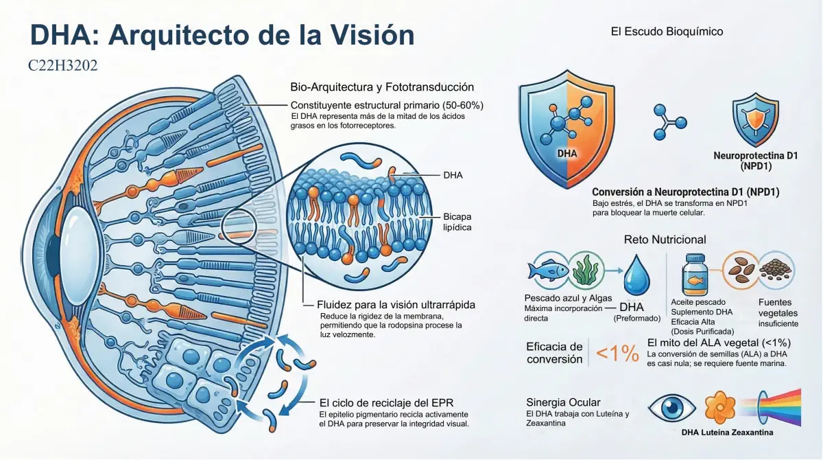
Download this infographic about the critical role of DHA in eye health in PDF format
Author:
Dr. Ronald Mauricio Sánchez Ávila, MD, PhD, MBA, MSc.
Ophthalmologist at Quirónsalud University Hospital and Olympia Center – Quirónsalud Group (Madrid).
Literature
- Age-Related Eye Disease Study 2 Research Group (2013). Lutein + zeaxanthin and omega-3 fatty acids for age-related macular degeneration: the Age-Related Eye Disease Study 2 (AREDS2) randomized clinical trial. JAMA, 309(19), 2005–2015. https://doi.org/10.1001/jama.2013.4997
- Swinkels, D., & Baes, M. (2023). The essential role of docosahexaenoic acid and its derivatives for retinal integrity. Pharmacology & Therapeutics, 247, 108440. https://doi.org/10.1016/j.pharmthera.2023.108440
- Shindou, H., et al. (2017). Docosahexaenoic acid preserves visual function by maintaining correct disc morphology in retinal photoreceptor cells. The Journal of Biological Chemistry, 292(29), 12054–12064. https://doi.org/10.1074/jbc.M117.790568
- Acar, N., et al. (2012). Lipid composition of the human eye: are red blood cells a good mirror of retinal and optic nerve fatty acids? PloS ONE, 7(4), e35102. https://doi.org/10.1371/journal.pone.0035102
- Birch, E. E., et al. (2010). The DIAMOND Study: a double-masked, randomized controlled clinical trial of the maturation of infant visual acuity as a function of the dietary level of docosahexaenoic acid. The American Journal of Clinical Nutrition, 91(4), 848–859. https://doi.org/10.3945/ajcn.2009.28557
- Mukherjee, P. K., et al. (2004). Neuroprotectin D1: a docosahexaenoic acid-derived docosatriene protects human retinal pigment epithelial cells from oxidative stress. PNAS, 101(22), 8491–8496. https://doi.org/10.1073/pnas.0402531101
- Bazan, N. G. (2009). Cellular and molecular events mediated by docosahexaenoic acid-derived neuroprotectin D1 signaling in photoreceptor cell survival and brain protection. Prostaglandins, Leukotrienes and Essential Fatty Acids, 81(2-3), 205–211. https://doi.org/10.1016/j.plefa.2009.05.024
- Arterburn, L. M., Hall, E. B., & Oken, H. (2006). Distribution, interconversion, and dose response of n-3 fatty acids in humans. The American Journal of Clinical Nutrition, 83(6 Suppl), 1467S–1476S. https://doi.org/10.1093/ajcn/83.6.1467S
- Brenna, J. T. (2002). Efficiency of conversion of alpha-linolenic acid to long chain n-3 fatty acids in man. Current Opinion in Clinical Nutrition and Metabolic Care, 5(2), 127–132. https://doi.org/10.1097/00075197-200203000-00002
- Bernstein, P. S., et al. (2016). Lutein, zeaxanthin, and meso-zeaxanthin: The basic and clinical science underlying carotenoid-based nutritional interventions against ocular disease. Progress in Retinal and Eye Research, 50, 34–66. https://doi.org/10.1016/j.preteyeres.2015.10.003
- Age-Related Eye Disease Study Research Group, SanGiovanni, J. P., et al. (2007). The relationship of dietary carotenoid and vitamin A, E, and C intake with age-related macular degeneration: AREDS Report No. 22. Archives of Ophthalmology, 125(9), 1225–1232. https://doi.org/10.1001/archopht.125.9.1225
Products related to DHA en la salud ocular
More entries at BrudyLab blog

Eye Health
Back to School and Children’s Vision

Maternal and Child
Breastfeeding and hydration

General
DHA and physical activity in summer: why the heat changes the rules

Artificial tears
Why is DHA so important for your eyes?
Eye Health
The critical importance of DHA in eye health
Eye Health
Glaucoma: Take care of your vision with nutrition and healthy habits

Maternal and Child
Benefits of breastfeeding
General