
Title: Why is DHA so important for your eyes?
Published: - Updated:
Author: Redacción BRUDYLAB
Reviewer: Dra. Leia Garrote - Medical Director
You’ve probably heard about Omega-3 in relation to the heart or brain. But there’s something you might not know: one of the places where Omega-3 is most crucial is in your eyes, and more specifically in the retina, the part of the eye that makes it possible for you to see.
Why is DHA so important for your eyes?
The great importance of DHA for your eyes
You’ve probably heard about Omega-3 in relation to the heart or brain.
But there’s something you might not know: one of the places where Omega-3 is most crucial is in your eyes, and more specifically in the retina, the part of the eye that allows you to see.
Omega 3 fatty acids are crucial for eyesight
The type of Omega-3 your retina needs has a name: DHA. It’s not interchangeable with other Omega-3s, and your body can’t produce enough of it on its own.
That’s why what you eat, or what you supplement, matters more than it seems.
Your retina is made, in large part, of DHA.
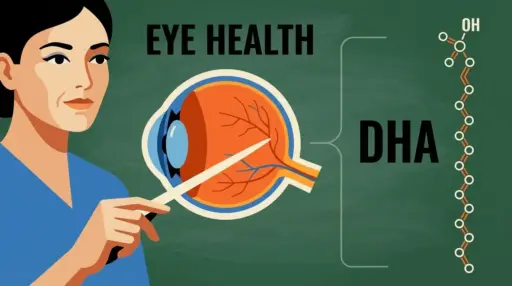
The retina is a very thin layer of tissue that lines the inside of the eye. It is responsible for capturing light and transforming it into signals that the brain can interpret as images. To do this, it needs special cells called photoreceptors, and these cells are made up of DHA in more than half of their membrane.
This is no coincidence. DHA has a molecular structure that allows it to be extraordinarily flexible, and this flexibility is precisely what photoreceptors need to react to light with the speed required for vision. When DHA levels drop, this flexibility is compromised, and with it, the quality of vision.
Think of it this way: if the retina were a muscle, DHA would be the component that allows it to stretch and contract without tearing. Without it, everything becomes stiffer and less efficient.
From before birth
DHA becomes critical even before birth. During the last trimester of pregnancy, the baby receives a massive transfer of DHA through the placenta. This supply is essential for the proper development of the retina and visual system.
Studies with infants have shown that DHA levels in the first months of life are directly related to the visual acuity the child will develop. This is no small detail: we are talking about the foundation upon which lifelong vision is built [1].
That’s why DHA is a particularly important nutrient during pregnancy and breastfeeding, and also in the diet of children in their early years.
Protect your eyes as you age
The retina works tirelessly. It is constantly exposed to light and generates a significant amount of waste products and free radicals—those unstable molecules that damage cells over time. To remain healthy, it needs a constant supply of nutrients to counteract this wear and tear.
DHA plays a role here that goes beyond being a simple structural component. When the retina is under stress, the body transforms some of the available DHA into a molecule called Neuroprotectin D1, which acts as an internal defense system: it slows the death of retinal cells, reduces inflammation, and helps maintain the central area of vision in good condition.
This protective capacity is especially relevant against Age-Related Macular Degeneration (AMD), the most common cause of vision loss in people over 60 in Western countries. It is estimated that by 2040, approximately 288 million people worldwide will be affected by some degree of this disease [2]. Maintaining adequate DHA levels throughout life is one of the most scientifically supported nutritional strategies for reducing this risk.
The problem with thinking that nuts and flax are enough
Here’s one of the most common misconceptions. Many people believe that by eating walnuts, flax seeds, or chia seeds, they are meeting their Omega-3 needs, including DHA. This is not the case.
These foods contain a type of Omega-3 called ALA, which is beneficial, but the body converts it into DHA very inefficiently. Several studies have measured this conversion, and the results are consistent: less than 1% of the ALA we ingest is ultimately transformed into DHA [3] [4]. The rest is used as energy or lost in the process.
Sources that do provide DHA directly include fatty fish such as salmon, sardines, mackerel, and herring, as well as certain shellfish. For those who don’t eat fish frequently, fish oil or microalgae supplements are the most evidence-based alternative.
The DHA does not work alone
A diet designed to protect eyesight isn’t limited to DHA. There are other molecules that work in a complementary way and together offer more complete protection:
- Lutein and zeaxanthin: two pigments present in green leafy vegetables and in orange and yellow foods that accumulate in the central part of the retina and act as a filter against blue light, the most harmful to photoreceptors.
- Vitamin E: an antioxidant that specifically protects the fats in retinal membranes, including DHA itself, from oxidative damage.
- Vitamin C: helps to keep the blood vessels inside the eye healthy.
- Zinc: a mineral necessary for the retina to properly use vitamin A and for the eye’s own antioxidant defense systems to function.
The AREDS2 study, one of the largest ever conducted on nutrition and eye health, showed that the combination of these nutrients significantly reduces the risk of AMD progressing to its more severe forms [2].
Frequently Asked Questions
At what age should I start worrying about DHA and my eyes?
Actually, DHA is relevant at all stages of life, although for different reasons. During pregnancy and early childhood, it helps build the retina. In adulthood, it helps maintain its health. And after age 50, it helps reduce the risk of degenerative diseases. There’s no age at which you should stop paying attention to it.
How much fish do I need to eat to meet my needs?
The usual recommendations suggest consuming oily fish at least twice a week. If this isn’t a regular practice, a fish oil or microalgae supplement may be a suitable alternative. In any case, for any specific eye health condition, it’s best to consult a healthcare professional.
Is plant-based Omega 3 not good for the eyes?
Plant-based omega-3 fatty acids (ALA) have benefits, but not directly for the retina. The conversion of ALA to DHA is so limited that it cannot be considered a reliable source for maintaining the levels that eye tissue needs. For eye health, marine sources of DHA are the ones with solid scientific support.
Do children also need DHA for their eyes?
Yes, and this is especially important in the first years of life. The retina continues to develop after birth, and DHA is an essential component of that process. A varied diet that includes oily fish, or appropriate supplementation when necessary, contributes to optimal development.
Glossary: Terms that appear in this article
- DHA (docosahexaenoic acid)
-
DHA is a special fat that belongs to the Omega 3 family. What makes it unique is that it is not primarily used as fuel, but as a building material: the body incorporates it directly into the membranes of the most active and demanding cells, especially in the brain, nerves, and retina of the eye.
When we talk about cell membranes, we’re referring to the outer layer that surrounds the cell and controls everything that enters and exits. For this layer to function properly, it needs to be flexible and responsive. DHA is precisely the type of fat that provides this flexibility: it has a long, curved molecular structure that prevents the membranes from stiffening.
The human body can produce a very small amount of DHA from other plant fats, but this amount is insufficient to meet the needs of the eyes and brain. Therefore, it is considered an essential nutrient that we must obtain from our diet, primarily from fatty fish such as salmon, sardines, or mackerel, or through fish oil or microalgae supplements.
It’s important not to confuse it with the Omega-3 found in walnuts or flaxseed oil. That type of Omega-3 (called ALA) has to undergo a series of transformations to become DHA, and the human body does this process very inefficiently. Therefore, eating walnuts is beneficial, but it’s not the same as consuming DHA directly.
- Retina
-
The retina is the innermost layer of the eye, attached to the back of the eyeball. If we think of the eye as a camera, the retina would be the sensor: the element that receives light, interprets it, and sends the information to the brain as an electrical signal.
The eye is composed of several types of cells, but the most important for vision are the photoreceptors. There are two types: rods, which allow us to see in low light and detect movement, and cones, which are responsible for sharp, color vision. Both types of photoreceptors have an extraordinarily high concentration of DHA in their membranes, much higher than any other tissue in the body.
The retina is a tissue that never rests: while we are awake, it is continuously processing information. This level of activity makes it especially vulnerable to wear and tear and damage accumulated over time, which explains why nutrition plays such a crucial role in its long-term maintenance.
The central area of the retina is called the macula, and it is responsible for the sharpest vision, the kind we use for reading, recognizing faces, or seeing details. It is also the area most affected by the disease known as Age-Related Macular Degeneration.
- Photoreceptors
-
Photoreceptors are the cells in the retina that capture light. Their name says it all: they are receptors of photons, the particles that make up light. When a photon of light reaches the eye and strikes the retina, these cells receive it and transform it into a signal that the optic nerve carries to the brain, where it is finally interpreted as an image.
There are two types. Rods are more numerous and more sensitive: they function in very low light and are what allow us to see at night or in dim light, although without color. Cones are less abundant but more specialized: they are concentrated in the center of the retina and are what give us color vision and the ability to distinguish fine details.
What makes these cells so interesting from a nutritional standpoint is their composition: more than half of the fats that make up their membranes are DHA. This high proportion is no coincidence. For a photoreceptor to function properly, it needs to respond to light in fractions of a second, and that’s only possible if its membranes are sufficiently fluid and flexible. DHA is what ensures this property.
When DHA levels are low, the membranes of photoreceptors become more rigid, their response to light slows down, and the quality of vision suffers. This effect is especially important in low-light situations and when viewing fine details.
- Taint
-
The Taint is a small area in the center of the retina, just a few millimeters across, responsible for the most precise and detailed vision. It’s the part of the eye we use when we look directly at something: reading text, seeing facial expressions, distinguishing the numbers on a clock, or threading a needle.
At the center of the macula is an even smaller area called the fovea, which is where the highest density of cones in the entire eye is concentrated. This makes it the point of maximum visual acuity.
The macula is also the area most susceptible to damage over time, precisely because it is the most active. Age-related macular degeneration specifically affects this area, causing a progressive loss of central vision that can become very debilitating, although in most cases peripheral vision is preserved.
Two pigments, lutein and zeaxanthin, accumulate naturally in the macula and act as a protective filter against high-energy light. Their presence in the diet, along with adequate levels of DHA, is one of the most studied nutritional factors related to long-term macular health.
- AMD (Age-Related Macular Degeneration)
-
Age-related macular degeneration (AMD) is an eye disease that affects the macula, the central area of the retina responsible for sharpest vision. Over time, the cells in this area deteriorate and stop functioning properly, leading to a progressive loss of central vision. Peripheral vision, which we use to see what is to the sides, is usually preserved, but the affected person gradually loses the ability to read, drive, or recognize faces.
It is the most common cause of severe vision loss in people over 60 in Western countries. There is no definitive cure, although treatments exist that can slow its progression in some cases.
There are two main forms. The dry form, which is more common, is characterized by a slow and progressive degeneration of the cells in the macula. The wet form, which is less common but more aggressive, involves the abnormal growth of blood vessels under the retina that can bleed and damage vision more rapidly.
Although age is the main risk factor, diet plays a significant role in both preventing and managing its progression. Regular consumption of DHA, lutein, zeaxanthin, vitamins C and E, and zinc is the nutritional intervention with the strongest scientific support in this area.
- Omega 3
-
Omega-3 fatty acids are a group of polyunsaturated fatty acids that the human body cannot produce on its own in sufficient quantities. This makes them essential nutrients that we must obtain from our diet.
Within the Omega-3 family, there are three main members. ALA is found in plant-based foods such as walnuts, flax seeds, and chia seeds. EPA and DHA are found primarily in fatty fish and shellfish. The difference between them is not only in their origin: they have distinct functions in the body. ALA is mainly used as an energy source. EPA has a significant anti-inflammatory effect and protects the cardiovascular system. DHA, as we have already seen, is a structural component of the brain and retina.
The body can convert ALA into EPA and DHA, but this process is so inefficient that it cannot rely on it to meet its needs. Therefore, while eating nuts or using flaxseed oil is beneficial, it is not a substitute for directly consuming oily fish or taking DHA and EPA supplements for those who need to ensure adequate levels of these nutrients.
- Neuroprotectin D1 (NPD1)
-
Neuroprotectin D1 is a molecule that the body itself produces from DHA when the retina is under stress, for example, when oxidative damage accumulates or when there is inflammation. Its name describes its function: it protects neurons and nerve cells, including those of the retina.
This molecule activates a series of defense mechanisms within the cells. It slows down the processes that lead to cell death, reduces local inflammation, and helps maintain the integrity of the macular area. In other words, when the eye is in trouble, the available DHA is converted into this compound, which acts as a rescue system.
This means that maintaining adequate DHA levels is not only important for building and maintaining photoreceptor membranes, but also for having a readily available reserve that the eye can draw upon when needed most. This is one of the strongest arguments in favor of a consistent and sustained DHA intake throughout life.
- Oxidative stress
-
Oxidative stress is the damage that occurs to cells when too many free radicals accumulate and the body doesn’t have enough antioxidants to neutralize them. Free radicals are unstable molecules generated as a byproduct of normal metabolism and also by external factors such as light exposure, pollution, or tobacco.
In the retina, oxidative stress is particularly intense. It’s a tissue that consumes a lot of oxygen, is continuously exposed to light, and has a very high concentration of polyunsaturated fats like DHA, which are especially vulnerable to oxidation. Therefore, the retina needs a very powerful antioxidant defense system to remain healthy.
When this system fails or when the damage exceeds the body’s repair capacity, retinal cells progressively deteriorate. This cumulative deterioration over the years is one of the main mechanisms behind diseases like age-related macular degeneration (AMD). Antioxidant nutrients, including DHA and its ability to generate Neuroprotectin D1, vitamins C and E, and macular pigments, are part of this defense system.
- Lutein and Zeaxanthin
-
Lutein and zeaxanthin are naturally occurring yellow and orange pigments belonging to the carotenoid group. They are found in foods such as spinach, kale, corn, yellow bell peppers, and squash. The body does not produce them on its own, so they must be obtained from the diet.
What makes them special in relation to eye health is that they selectively accumulate in the macula, the central area of the retina. There, they form a kind of filter that absorbs high-energy blue light before it reaches the photoreceptors and damages them. It’s as if the eye has its own internal sunglasses.
In addition to this filtering effect, they act as antioxidants directly in the retinal tissue, neutralizing free radicals. Several studies, including AREDS2, have shown that maintaining adequate levels of lutein and zeaxanthin significantly reduces the risk of AMD progressing to its more severe forms.
A diet rich in leafy green vegetables and brightly colored foods (orange, yellow) is the best way to ensure an adequate intake of these pigments. In some cases, supplementation may be a complementary option, especially for people at higher risk of macular degeneration.
- Phototransduction
-
Phototransduction is the process by which the eye converts light into an electrical signal that the brain can interpret. It is, essentially, the mechanism that makes vision possible.
When light enters the eye and reaches the retina, it strikes the photoreceptors. Inside each photoreceptor is a protein called rhodopsin, which reacts to light by changing shape. This change in shape triggers a series of chain reactions that ultimately generate an electrical signal. This signal travels along the optic nerve to the brain, which interprets it and converts it into the image we see.
This entire process occurs in fractions of a second, and for it to be possible, the photoreceptor membranes must be highly fluid and flexible. If they were rigid, rhodopsin wouldn’t be able to change shape quickly enough. This is where DHA comes in: it’s the component that ensures this fluidity. Without adequate levels of DHA, the phototransduction process slows down, and the quality of vision, especially in low light, deteriorates.
- AREDS2 Study
-
The AREDS2 (Age-Related Eye Disease Study 2) is one of the largest and most rigorous clinical trials ever conducted on nutrition and eye health. It was coordinated by the U.S. National Eye Institute and included more than 4,000 participants followed for several years.
Their goal was to determine if certain nutritional supplements could slow the progression of age-related macular degeneration. The results showed that a specific combination of lutein, zeaxanthin, vitamins C and E, and zinc significantly reduced the risk of the disease progressing to its more severe forms.
One of the most important findings of the study was that replacing beta-carotene (present in the original formula of the previous study, AREDS1) with lutein and zeaxanthin improved the results and eliminated a risk associated with beta-carotene in smokers. Since then, the AREDS2 combination has been considered the gold standard in nutritional supplementation for macular health.
The study also analyzed the role of Omega-3 fatty acids, although in that case the results did not show a statistically significant benefit when taken in combination with the other nutrients. This does not invalidate the accumulated evidence on DHA and the retina from other studies, but rather reflects the complexity of isolating the effect of a specific nutrient in a multifactorial disease.
- Retinal Pigment Epithelium (RPE)
-
The retinal pigment epithelium is a layer of cells located just behind the photoreceptors. Although less well-known than the retina as a whole, it performs essential functions for maintaining good vision.
Its primary function is to maintain the photoreceptors. Every day, the photoreceptors renew parts of their internal structures, and the discarded fragments are phagocytosed—literally engulfed and recycled by the RPE cells. This process removes waste products that, if they accumulated, would damage the retina, and recovers valuable components, including DHA, which are returned to the photoreceptors for reuse.
The RPE is also responsible for producing Neuroprotectin D1 from DHA when the retina is under stress. In a sense, it’s the eye’s defense lab: it detects the threat and produces the antidote.
When the retinal pigment epithelium (RPE) begins to fail, due to age-related wear and tear or factors such as smoking or excessive exposure to light without protection, debris accumulates under the retina, forming deposits known as drusen. These drusen are an early warning sign of age-related macular degeneration (AMD) and one of the first things ophthalmologists look for during eye exams for people over 50.
Literature
- Birch, E. E., et al. (2010). The DIAMOND Study: a double-masked, randomized controlled clinical trial of the maturation of infant visual acuity as a function of the dietary level of docosahexaenoic acid. The American Journal of Clinical Nutrition, 91(4), 848–859. https://doi.org/10.3945/ajcn.2009.28557
- Age-Related Eye Disease Study 2 Research Group (2013). Lutein + zeaxanthin and Omega 3 fatty acids for age-related macular degeneration: the Age-Related Eye Disease Study 2 (AREDS2) randomized clinical trial. JAMA, 309(19), 2005–2015. https://doi.org/10.1001/jama.2013.4997
- Brenna, J. T. (2002). Efficiency of conversion of alpha-linolenic acid to long chain n-3 fatty acids in man. Current Opinion in Clinical Nutrition and Metabolic Care, 5(2), 127–132. https://doi.org/10.1097/00075197-200203000-00002
- Arterburn, L. M., Hall, E. B., & Oken, H. (2006). Distribution, interconversion, and dose response of n-3 fatty acids in humans. The American Journal of Clinical Nutrition, 83(6 Suppl), 1467S–1476S. https://doi.org/10.1093/ajcn/83.6.1467S
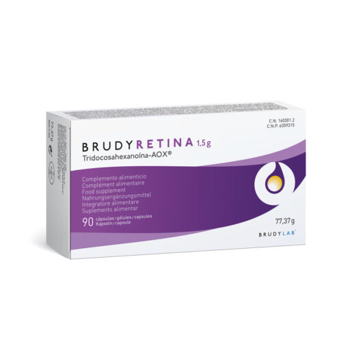
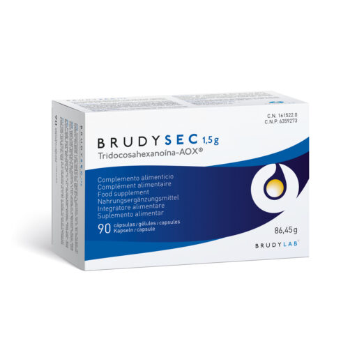
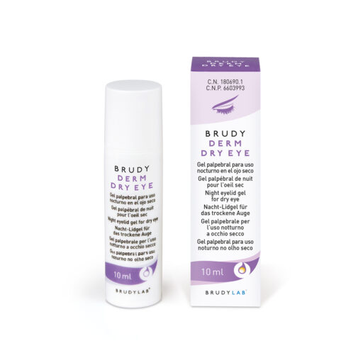
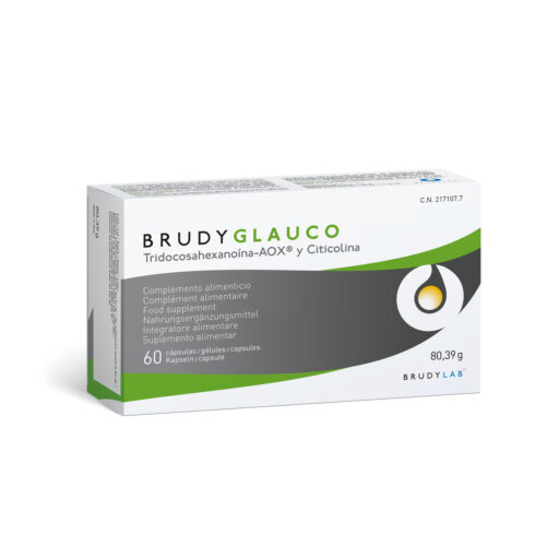
Products related to DHA para la vista
More entries at BrudyLab blog

Eye Health
Back to School and Children’s Vision

Maternal and Child
Breastfeeding and hydration

General
DHA and physical activity in summer: why the heat changes the rules

Artificial tears
Why is DHA so important for your eyes?
Eye Health
The critical importance of DHA in eye health

Eye Health
Glaucoma: Take care of your vision with nutrition and healthy habits

Maternal and Child
Benefits of breastfeeding
General